Figure 3.

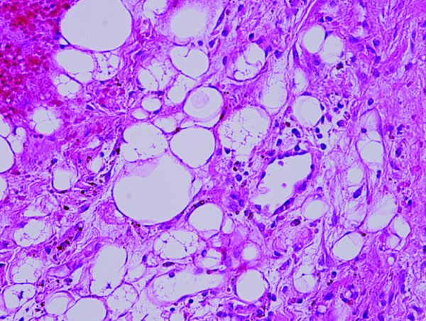
Microscopic findings revealing polygonal cells with eosinophilic cytoplasms within a prominent vascular component (the typical zellballen structures, H&E 200×).

Microscopic findings revealing polygonal cells with eosinophilic cytoplasms within a prominent vascular component (the typical zellballen structures, H&E 200×).