
Fig. 3.
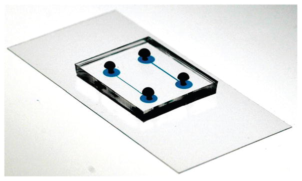
Picture of the microfluidic device. The device consists of two layers: a layer of PDMS containing the channels bonded to a glass slide covered with PDMS. The length of one channel is 1 cm and the channels were filled with blue dye to make them easy to visualize.