
Figure 1.
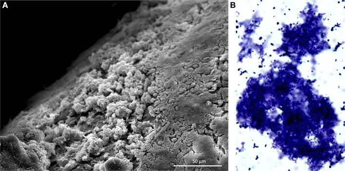
Pneumococcal biofilms form in the nasopharynx. (A) Scanning electron microscopy image of S. pneumoniae biofilms formed on the nasal septum of a mouse. Mice were experimentally colonized 7 days prior. Biofilms are the non-contiguous aggregates on the left. (B) S. pneumoniae biofilm aggregate in nasopharyngeal lavage fluid. Sample was collected from mouse 14 days after experimental colonization. Pneumococci were stained with crystal violet and visualized with a light microscope at 400X. Image credit: Krystle Blanchette.