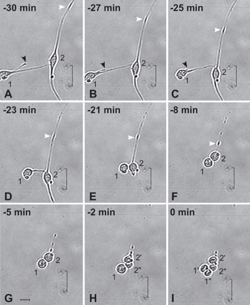
Fig. 2.
Anterior subventricular zone (SVZa) neuronal progenitor cells retract their processes prior to cytokinesis. A 30 min series of time-lapse videomicroscopic photomicrographs of two SVZa neuronal progenitor cells beginning 5 DIV. The time that Cell no. 1 undergoes cytokinesis is considered t = 0. The position of each cell is visualized relative to marks on the underside of the growing surface (indicated by a bracket). The behavior of Cell no. 1 is described first and then Cell no. 2. (i) Cell no. 1. (A–C) At t = −30 min (A), Cell no. 1 has relatively long processes. Between 30 and 25 min prior to cytokinesis, Cell no. 1 retracts its shorter process (black arrowhead). (C–E) Between 25 and 21 min prior to cytokinesis, the longer process of Cell no. 1 (the process contacting Cell no. 2) retracts. Concurrently, the soma of Cell no. 1 moves towards Cell no. 2. (C–F) Cell no. 1 rounds up. (G and H) Cell no. 1 remains in contact with Cell no. 2 and, after Cell no. 2 divides, with its progeny. (I) At t = 0 min, Cell no. 1 divides, generating two daughter cells (labeled 1′ and 1″). (ii) Cell no. 2. (A–G) From t = −30 to t = −5 min, Cell no. 2 retracts both of its processes. Note that the shorter process (below the soma) retracts more quickly (see E) than the longer process (above the soma). The longer process is in contact with the process of another cell (the soma of which is located beyond the edge of the image at the upper right) giving the appearance of a single long process. The change in the position of an enlarged dense segment of the longer process of Cell no. 2 (white arrowhead in A–F) is indicative of the retraction of the process towards its soma. While Cell no. 2 is withdrawing its processes, its somatic morphology is transforming from elongated (A–C) to round (D–G). (H and I) At t = −2 min, Cell no. 2 undergoes cytokinesis, generating two daughter cells (labeled 2′ and 2″). Note that, although Cell no. 1 completes the retraction of its processes before Cell no. 2 (D–F), Cell no. 2 divides first (compare H with I). The alignment of the chromosomes is evident in the daughter cells of both Cell no. 1 (1′ and 1‴) and Cell no. 2 (2′ and 2″). See also supplementary Video S1, which shows the dynamics of process retraction of other cells. Scale bar, 20 µm in G (applies to all panels).

