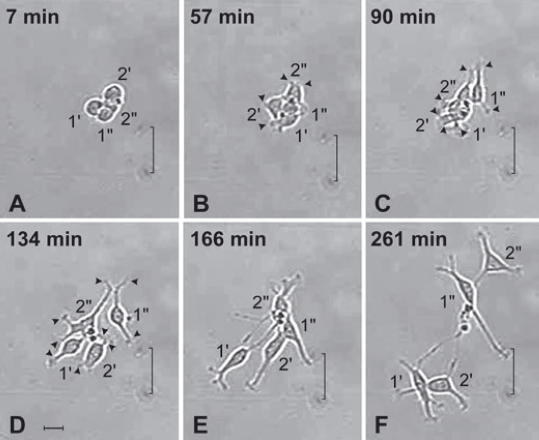
Fig. 3.
Anterior subventricular zone (SVZa) progenitor cells extend processes following cytokinesis. A series of time-lapse videomicroscopic photomicrographs of the four daughters of Cell nos 1 and 2 (continued from Fig. 2I) as they elaborate processes during the first 4.5 h following their generation. Time is measured relative to t = 0 in Fig. 2I. The position of each cell is visualized relative to marks on the underside of the growing surface (indicated by a bracket). (A) Following cytokinesis, the four newly generated cells are clustered and lack processes. (B and C) The newly generated cells initiate process extension at nearly the same time. All four cells have short processes ending in an active, broad growth cone-like structure (e.g. arrowheads). Most cells have two or more processes. (D) The cells begin to disperse by t = 134 min. At this time most of the cells have long processes (e.g. arrowheads). (E and F) The cells continue to separate from each other, form new processes and lengthen their existing processes. Together the panels of this figure demonstrate that newly generated SVZa progenitor cells begin to extend elaborate processes within a short time after division. See also supplementary Video S1, which shows the dynamics of process extension of other cells. Scale bar, 20 µm in D (applies to all panels).

