
Figure 2.
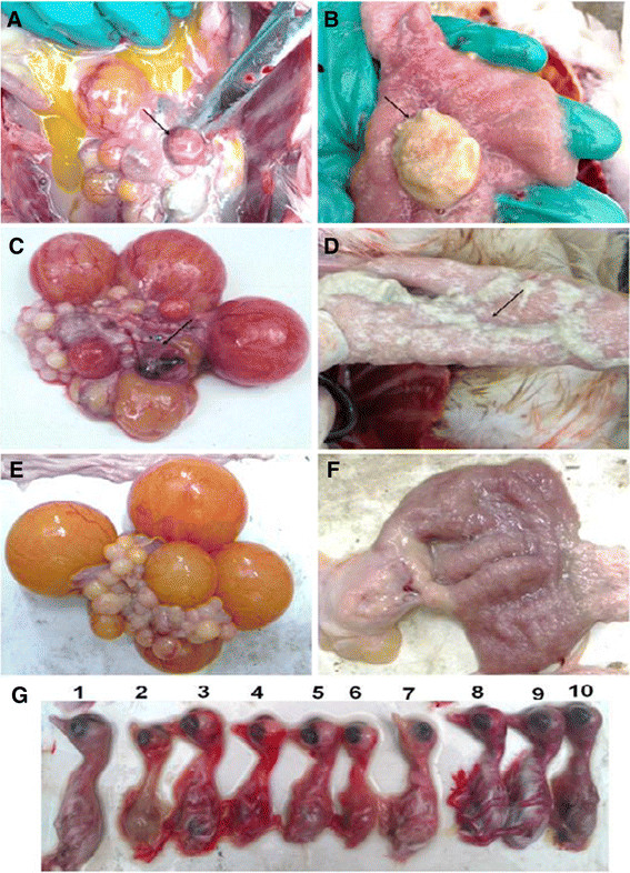
Pathological studies of the infected ducks and the inoculated duck embryos. i. The naturally infected ducks were euthanized and anatomic investigations show the prevalence of white or yellow discharge in uterine (B), ovarian hemorrhage and abdominal egg yolk accumulated (A), and uterine flushing (A). The artificial infected laying ducks experiencing necropsies revealed white discharge in the uterine (D) and ovarian hemorrhage (C). Mock duck uterine (F) and ovarian (E). ii. Duck embryos, 5–7 days post inoculation, growth retardation appeared (G-2, −4, −5, −6), haemorrhage (G-3, −4, −5, −6, −8, −9, −10), and death (G-2, −3, −4, −5, −6, −7, −8, −9, −10), Control (G-1).