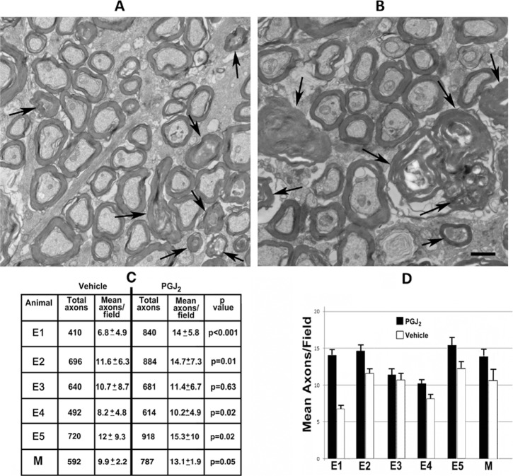
Figure 9.
Electron microscopic analysis of ONs of post pNAION vehicle-injected and PGJ2-treated eyes (n = 5). (A, B) Representative TEMs of animal E5. (A) Vehicle-injected ON. (B) PGJ2-treated ON. There is relative reduced axon packing density in the ON of the vehicle-injected eye compared with the PGJ2-treated eye. Although degenerating axons are seen in both ONs (arrowheads), there is increased interaxonal debris in the vehicle-injected nerve (compare [A] and [B]), and the myelin sheaths are thinner in the nerve from the vehicle-injected eye than in the ON from the PGJ2-treated eye. Demyelinated axons are seen in both nerves (arrowheads). (C) ON axon quantification in vehicle-injected and PGJ2-treated eyes. Sixty axon fields were randomly selected in each ON, and axons counted in each 135.5-um2 field, using a stereological frame (see Methods). Total counted axons are indicated in the first (vehicle-injected) and third (PGJ2-treated) columns. Mean axons/field are indicated in the second (vehicle-injected) and fourth (PGJ2-treated) columns. Significance between axon counts was determined using a two-tailed t-test for individual animals and Wilcoxon Rank Sum test for the group as a whole. PGJ2-treated eyes had more preserved axons than vehicle-injected eyes in all animals. The difference was significant in four animals (E1: P < 0.001; E2: P = 0.01; E4: P = 0.02; E5: P = 0.02) as well as for the group as a whole (P = 0.05). (D) Bar graph showing relative axon preservation in the five individual test animals as well as for the group as a whole (for vehicle-injected eyes, SD = 2.04 and SE = 1.01; for PGJ2-treated eyes, SD = 3.84 and SE = 1.34). Scale bar: 500 nm. Error bars are ±2 SE.

