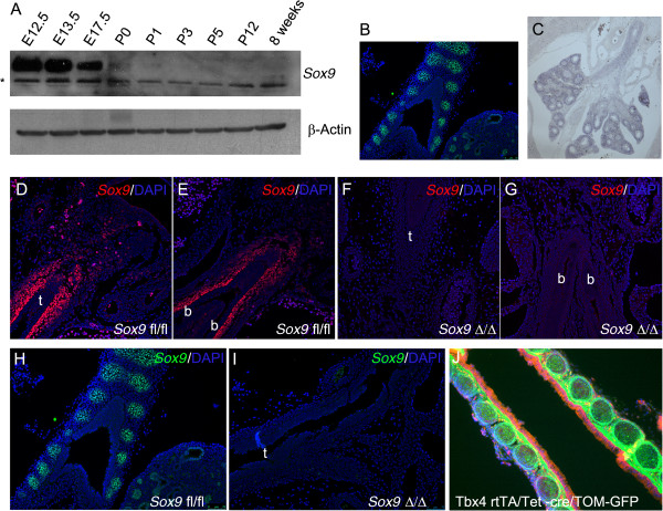
Figure 1.

Expression of Sox9 during lung development. (A) Western blot analysis of Sox9 protein expression during lung development at different stages (*non-specific bands). (B)Sox9 protein expression at E15.5 stage: immunofluorescence localization of Sox9 showed expression of Sox9 by tracheal immature cartilage and distal lung epithelium. (C) Expression pattern of endogenous Tbx4 gene in E15.5 lung: Tbx4 is expressed by tracheal mesenchyme and by most, but not all of the distal lung mesenchyme. (D-I)Sox9 immunostaining, showing effective deletion of mesenchyme Sox-9 expression in the mutant lungs at embryonic day (D-G) E12.5 and (H, I) E15.5 stage. (J) Cell tracing using Tbx4-rtTA line. Tbx4-rtTA/Tet-on-cre males were bred with mT/mG females. E7.5 staged pregnant females were fed with doxocycline, and E18.5 embryos were collected. Lung epithelial cells expressed red fluorescent protein, while the mesenchyme cells are green because they are derived from Tbx4-expressing cells (t, trachea; b, bronchus).
