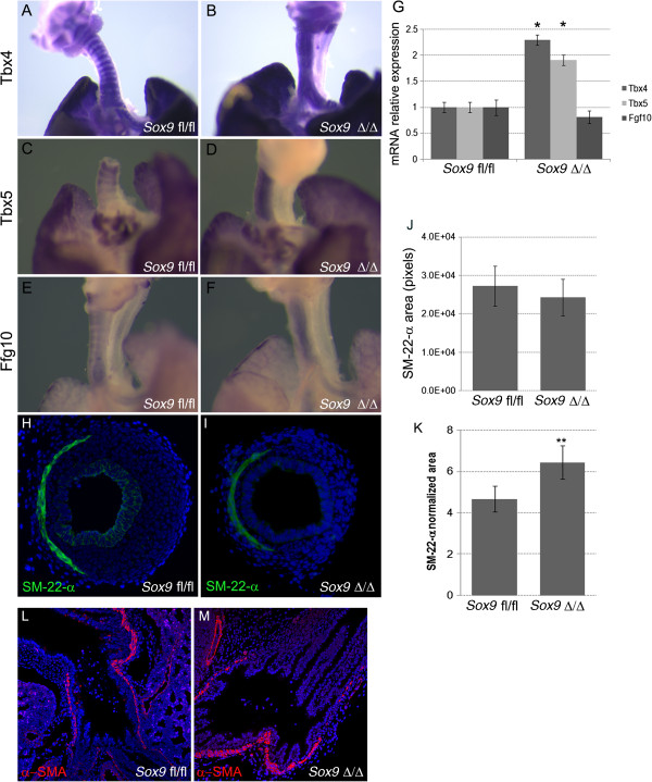
Figure 5.

Tbx4, Tbx5, and Fgf10 in situ hybridization results.In situ hybridization for (A, B)Tbx4 and (C, D)Tbx5 was performed on lung at embryonic day (E) 15.5 to identify changes in expression patterns. Tbx4 andTbx5 were expressed by the mesenchyme surrounding the tracheal rings in the normal control embryonic lung tracheas (A, C), whereas Sox9 knockout lungs displayed diffuse expression of (B)Tbx4 and (D)Tbx5 along all the tracheal mesenchyme. (E, F) The Fgf10 expression pattern was lost in the Sox9Δ/Δ trachea. (G) Real time RT-qPCR PCR was used to determine whether expression of Tbx4 and Tbx5 mRNA was increased in the mutant mouse trachea versus wild-type mouse trachea (*P < 0.05). Fgf10 expression was not significantly different between by Sox9Δ/Δ vs. the Sox9fl/fl tracheas. (H-K) Trachealis smooth muscle cells did not change after Sox9 knockout. Staining for Sm-22-α was used to identify the trachealis smooth muscle cells on transverse sections of normal and transgenic mouse tracheas. (J, K) Even though the absolute area of trachealis smooth muscle cells did not change, the Sm-22-α relative area was significantly higher in the Sox9Δ/Δ(J) compared with (K) the Sox9fl/fl trachea, because of the smaller transversal area of the mutant trachea. **P = 0.015. Values are mean ± SD, n = 5. (L, M). Staining for α-smooth muscle actin (α-SMA) was used to highlight smooth muscle cells. Sox9 deletion did not affect lung smooth muscle cell proliferation or differentiation.
