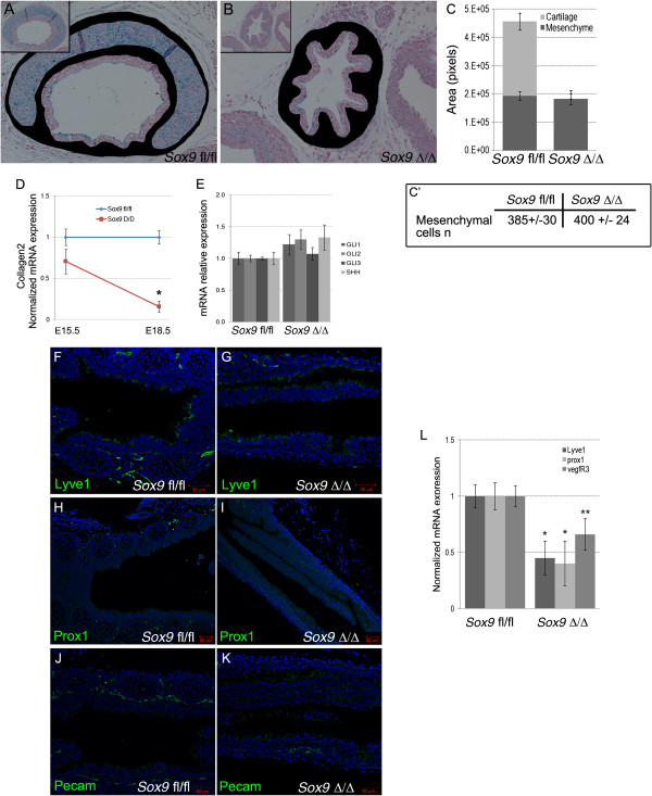
Figure 9.

Lymphatic vascular system was affected by Sox9 deletion. (A-C) Tracheal mesenchyme area quantification analysis did not reveal changes between the wild-type and mutant tracheas (tracheal mesenchyme was stained in black). (C′) The number of mesenchymal cells per section was similar between wild-type and mutant trachea. (D, E) Real time RT-qPCR for Collagen2, Gli1, and Shh on mRNA from wild-type and mutant tracheas at embryonic day (E) 15.5. Col2 expression was decreased in the Sox9Δ/Δ trachea starting at E15.5 stage. No statistically significant differences in expression of Shh, Gli1, Gli2, or Gli3 was observed between Sox9fl/fl and Sox9Δ/Δ. (F-L) The lymphatic vascular system was underdeveloped in the mutant trachea. Immunostaining of the lymphatic markers (F, G) Lyve1, (H, I) Prox1, and (J, K) Pecam revealed that Sox9 deletion resulted in impaired lymphatic vessel formation. (L) Real time RT-qPCR for Prox, Lyve1, and Vegfr3 confirmed that the lymphatic vascular system was not properly developed in the Sox9 knockout tracheas. *P < 0.05, **P = 0.08.
