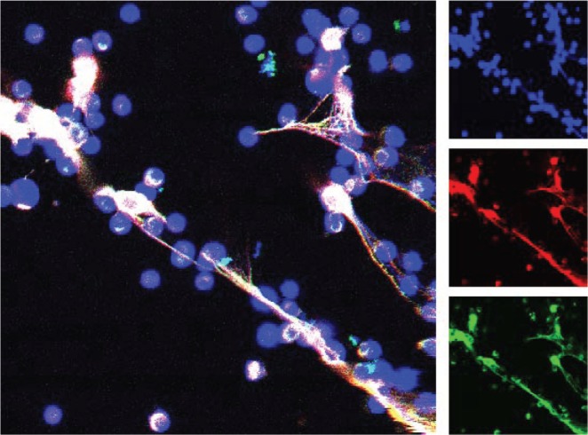
Figure 2.
Detection of histone H1 on neutrophil extracellular chromatin “traps”. Human neutrophils were purified and incubated with A23187 ionophore for 2 hours before processing for confocal microscopy with anti-linker histone H1 antibody. The main image shows three color confocal laser scanning micrograph that combines staining of DNA (blue) and anti-H1 antibody (detected by two different secondary antibodies, shown in green and red). Linker histone H1 remains associated with neutrophil extracellular traps.

