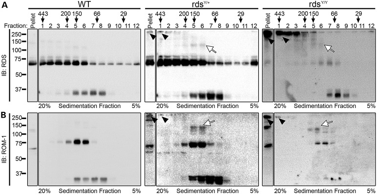
Figure 7.
Y141C-RDS leads to formation of abnormal high-molecular-weight oligomers. To analyze RDS/ROM-1 complexes under non-denaturing conditions, whole retinal extracts were prepared and run on a continuous 20% (fraction 1) to 5% (fraction 12) non-reducing sucrose gradient. Fractions were collected and analyzed using non-reducing SDS–PAGE/western blotting with antibodies specific for RDS (top) and ROM-1 (bottom). The position that molecular weight markers appear when using this protocol is listed above the fraction number for reference. In the rdsY/+ a full complement of normal RDS monomer and dimer were seen; however, abnormal high-molecular-weight complexes are observed in the heavier fractions as well as the pellet (arrowheads, middle). The rdsY/Y lacks normal higher-order complexes (dimers in high-density fractions) of RDS which are replaced by very distinct high-molecular-weight complexes in fractions 1–3 and in the pellet (arrowheads, right). N = 3–4 independent experiments/genotype.

