Figure 3.
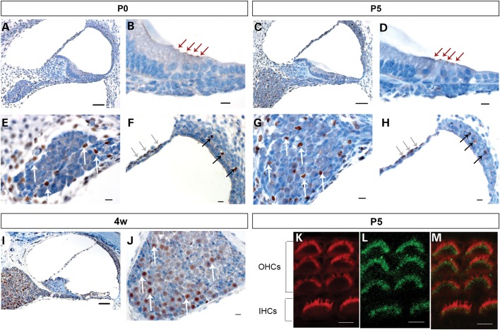
Sik3 is expressed in cochlear structures at different developmental stages. Mouse cochlear sections were stained with primary Sik3 antibody (brown) and counterstained with haematoxylin (blue). A, B, E and F: at the day of birth (P0), Sik3 expression was detected in the apex of inner and outer hair cells (B, red arrows), in the perilymph-facing layer of the Reissner's membrane (F, grey arrows), near blood vessels of the intermediate layer of the stria vascularis (F, black arrows), as well as in cells of the spiral ganglion (E, white arrows) and cells surrounding the ganglion. C, D, G and H: at 5 days postnatal (P5), Sik3 expression was found in the hair cells (D, red arrows) and small cells of the spiral ganglion (G, white arrows). I and J: at 4 weeks postnatal, Sik3 expression remained in the spiral ganglion (J, white arrows), Reissner's membrane and stria vascularis but could not be detected in the apex of the hair cells (I). K, L and M: confocal imaging of Sik3 expression in the stereocilia at 5 days postnatal (L, green), compared with Phalloidin expression (K, red) showed that Sik3 was expressed in the region around the base of the stereocilia (M, merged image of K and L). IHCs, inner hair cells; OHCs, outer hair cells. Scale bars: A, C and I: 50 µm; B, D, E, F, G, H and J: 10 µm; K, L and M: 5 µm.

