
Figure 2.
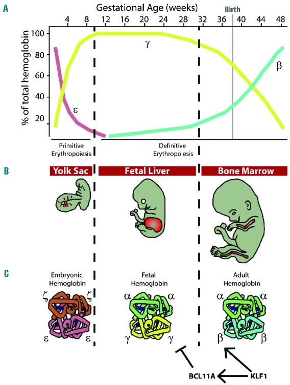
Human erythropoiesis and developmental pattern of hemoglobin expression. (A) Hemoglobin switching in man. Around week 6 of gestation, embryonic globin (ε) is silenced and fetal globin (γ) starts to be expressed. Perinatally the switch to adult globin (β) occurs. For the α-like globins, a single switch from the embryonic (ζ) to adult (α) globin occurs (not shown). (B) Major anatomical sites of hematopoiesis during development. Erythropoiesis occurs in the blood islands of the yolk sac in the first 8 weeks of gestation, then in the fetal liver between 8 and 32 weeks, and finally in the bone marrow from 32 weeks on. Around birth the spleen serves as a transient erythropoietic organ (not shown). (C) Structure of the main human hemoglobins expressed during development. Embryonic globin (ζ2ε2; HbE Gower-1); fetal hemoglobin (α2γ2; HbF) and adult hemoglobin (α2β2, HbA). KLF1 and BCL11A are two transcription factors with key roles in the developmental regulation of hemoglobin expression. In particular, KLF1 activates β-globin and BCL11A, which in turn represses γ-globin expression.