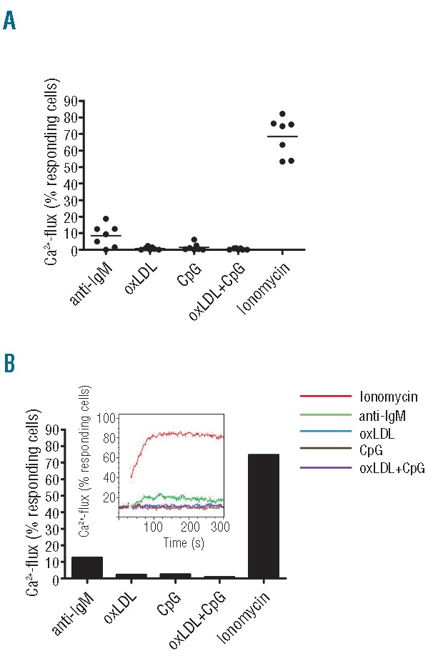
Figure 2.

oxLDL ligation does not induce Ca2+-flux. (A) Subset #1 lymphocytes from 7 CLL cases were analyzed for Ca2+-release. Cells were loaded with the Ca2+-sensitive dye Fluo-4-AM and analyzed by flow cytometry before and after addition of oxLDL (50 μg/mL), anti-IgM F(ab´)2 (20 μg/mL), and/or CpG (10 μg/mL). The calcium ionophore ionomycin was used as a positive control. The dot plot shows percent of responding cells of total viable cells. (B) The Ca2+-mobilization data of CLL2 (1 of the 7 CLL cases included in A) is shown for clarity. The diagrams show percent responding cells of total viable cells. The insert shows flow image for Ca2+-flux responses. (See Online Supplementary Figure S2 for all flow images).
