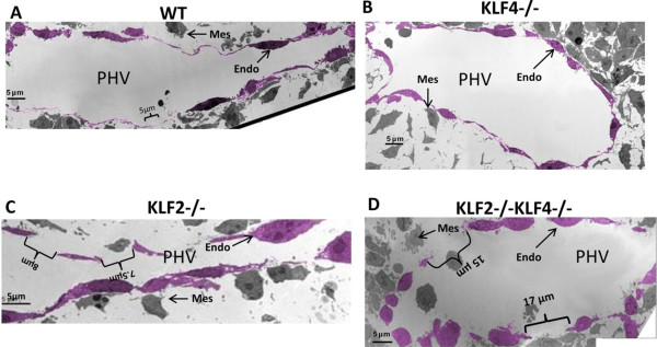
Figure 3.

Electron micrographs confirm disruption of the endothelial layer of the primary head vein in E9.5 KLF2-/-KLF4-/- embryos. Images were taken of the PHV at the level of the optic vesicle. The endothelial cell layer was pseudo-colored using GIMP (GNU Image Manipulation Program) version 2.6 open-source digital photo editing software. A) WT embryo (n = 1) has a continuous endothelial layer with slight gaps less than 5 μm in length. B) Only slight gaps were observed in KLF4-/- embryo (n = 1). C) One of the two KLF2-/- embryos presented a few gaps of 8 μm and 7.5 μm in length. D) KLF2-/-KLF4-/- embryos (n = 2) had consistent disruptions of the endothelial membrane with gaps as long as 17 μm. PHV: primary head vein lumen: Endo: endothelial cell; Mes: mesenchymal cell.
