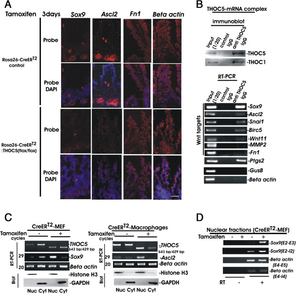
Figure 6.

Wnt signaling downstream genes are THOC5 target mRNAs. (A)In situ hybridization: Frozen sections of small intestine from Rosa26-CreERT2:THOC5 (flox/flox) or Rosa26-CreERT2 mice 3 days after tamoxifen treatment were hybridized with Sox9, Ascl2, Fn1 or beta-actin specific probes and counterstained with DAPI. Bars represent 40 μm. (B) Endogenous THOC5 was immunoprecipitated from the nuclear fraction of mouse embryonic fibroblasts or mouse hepatocellular carcinoma cell line HEPA1-6 using THOC5 specific monoclonal antibody (anti THOC5 IgG) and control mouse IgG (control IgG). THOC5-mRNA complex was analyzed by THOC5 or THOC1 specific immunoblot or bound RNAs were examined by Sox9, Ascl2, Snai1, Birc5, Wnt11, MMP2, Fn1, Ptgs2, GusB, and beta-actin specific RT-PCR. (C) MEF or Bone marrow derived macrophages were isolated from Rosa26-CreERT2:THOC5 (flox/flox) mice. Cells were incubated with (+) and without (−) tamoxifen (10 μM) for three days, and nuclear (Nuc) and cytoplasmic (Cyt) RNAs were then isolated and applied to spliced Ascl2, Sox9, THOC5 or actin specific semi-quantitative RT-PCR using primers as described in Table 1. cDNA from all samples were standardized by adjusting equal levels of actin mRNA in both fractions. Protein extracts were supplied for GAPDH and Histone H3 specific immunoblot (Blot). We performed 3 independent experiments and we show one example of representative data. (D) Aliquots of nuclear RNAs in (C) were supplied for spliced (E2-E3: Exon2-Exon3) or unspliced(E2-I2: Exon2-Intron2) Sox9, or spliced (E4-E5: Exon4-Exon5) and unspliced (E4-I5: Exon4-Intron4) beta actin specific RT-PCR (primers: Table 1). RT: reverse transcriptase reaction.
