
Figure 1.
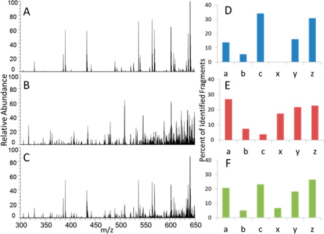
MS/MS spectra of ubiquitin (13+). (A) ETD (15 ms in HCD cell), (B) UVPD (one pulse, 2.5 mJ in HCD cell), (C) ETUVPD (15 ms ETD in HCD cell followed by UVPD using one pulse 2.5 mJ in HCD cell), and corresponding distribution of ion types in panels D, E, and F. All spectra are shown on the same scale.