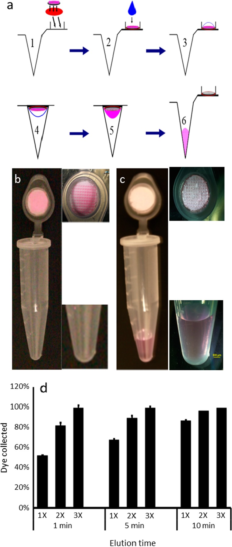
Figure 2.

Collection of analytes from the microneedle patch by centrifugation: (a) A microneedle patch containing analytes of interest is adhered to the inner surface of a microcentrifuge tube cap (1). A drop of water is dispensed to the microneedles (2), allowed to extract/dissolve analytes from the microneedles (3), and the cap is closed (4). The analytes are then centrifuged to draw the analyte solution from the cap to the base of the microcentrifuge tube (5), which can then be removed from the bottom of the tube for subsequent analysis (6). This method is illustrated by showing a microneedle patch containing pink dye (sulforhodamine), which serves as a model analyte, before (b) and after (c) application of water and centrifugation. The results are quantified in part d, which shows the percentage of analyte collected from the microneedles into the elution fluid after application of 100 μL of water for 1, 5, or 10 min, and centrifugation at 300g, which was carried out one (1×), two (2×), or three (3×) times (n = 5 replicates ± standard deviation error bars).
