Figure 4.
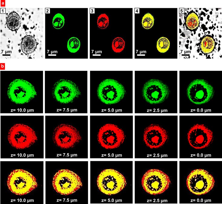
Intracellular localization of anisotropic biodegradable polymer/lipid Janus nanoparticles. (a) Representative images of A549 human lung cancer cells incubated 24 h with nanoparticles: 1, Light; 2–4, Fluorescence; 2, Polymeric (PLGA) phase of nanoparticles was labeled with FITC (green fluorescence); 3, Lipid (precirol) phase was labeled with DiR (red fluorescence); 4, Superimposition of green and red fluorescence images shows colocalization of PLGA and lipid phases of nanoparticles (yellow color); and 5, Superimposition of light, green and red fluorescence images shows intracellular localization of nanoparticles. (b) Representative confocal microscopy (z-series, from the top of the cell to the bottom) images of A549 human lung cancer cells incubated for 24 h with anisotropic biodegradable polymer/lipid Janus nanoparticles. Polymeric (PLGA) phase of nanoparticles was labeled with FITC (green fluorescence); lipid (precirol) phase was labeled with DiR (red fluorescence). Superimposition of green and red fluorescence images shows colocalization of PLGA and lipid phases of nanoparticles (yellow color).

