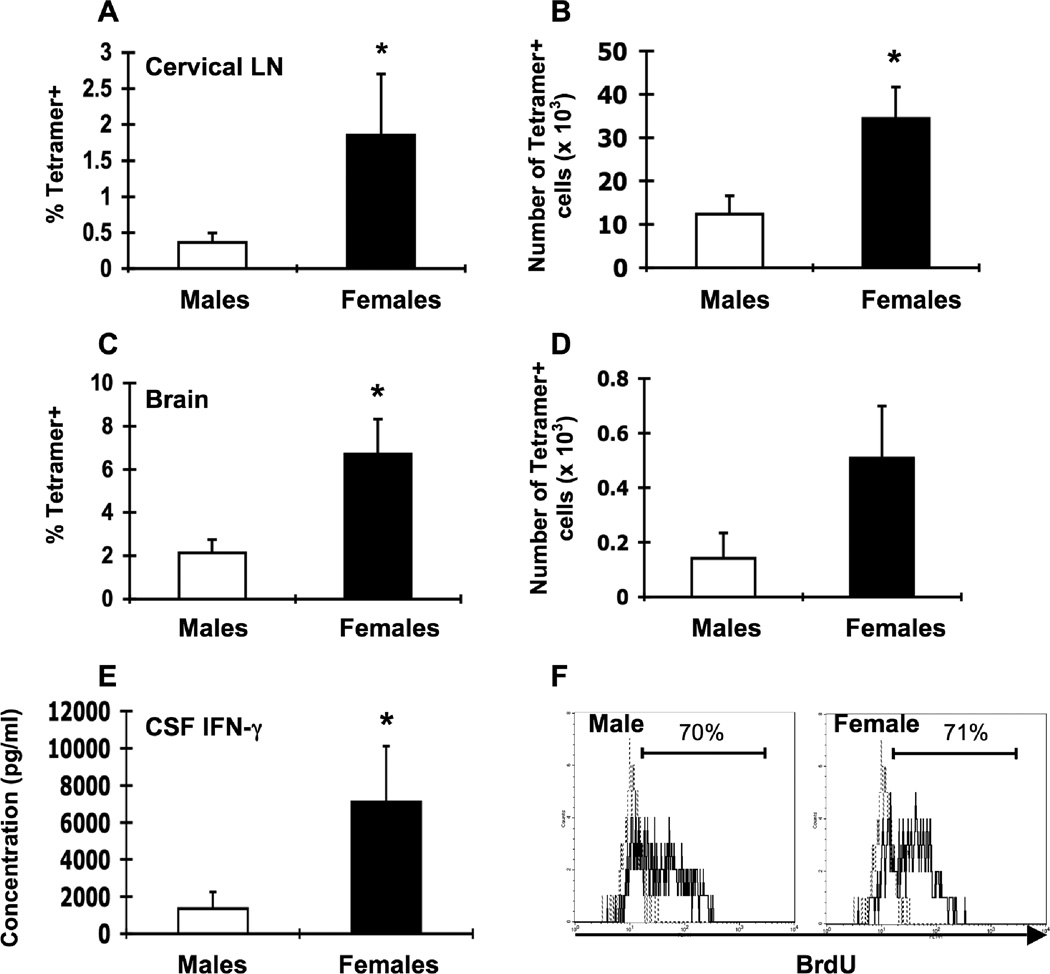
Figure 2.
Lower LCMV-specific T cell responses in male β2m−/− mice. Groups of male (white bars, n = 5–7) and female (black bars, n = 5–7) β2m−/− mice were infected i.c. with 1 × 10³ pfu LCMV, sacrificed eight days later, and perfused with 10 ml PBS. Mononuclear cells from cervical LN and brains were isolated, stained with I-Ab gp61-80 tetramer, and analyzed by flow cytometry. Percentages (A, C) and total numbers (B, D) of tetramer-positive cells in the cervical LN (A, B) and brain (C, D) or infected males and infected females. (E) IFN-γ levels in the CSF. Data represent the mean ± SEM and are pooled from two independent experiments and are representative of at least three independent experiments with similar results. *p ≤ 0.05 (Mann-Whitney test, Minitab for Windows). (F) Groups of male and female β2m−/− mice were infected i.c. with 1 × 10³ pfu LCMV. Mice received i.p. injections of 1 mg BrdU on days 4–7 after infection and proliferation of LCMV-specific T cells was assessed on day 8 by I-Ab gp61-80 tetramer staining and intracellular BrdU staining and flow cytometric analysis. Representative histograms show the frequency of CD4+ I-Ab gp61-80 gated cells that were BrdU+ in cervical LN from infected male (left) and female (right) mice (solid lines). Dotted lines represent BrdU staining in an infected mouse that did not receive BrdU injections.

