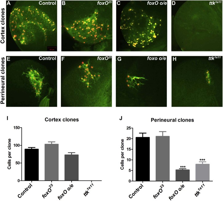
Fig. 4.
foxO and ttk69 regulate glial proliferation in the postembryonic brain. (A–D) Representative repo-MARCM cortex clones marked with GFP (green) and nuclear-RFP (red) expression. (E–H) Representative repo-MARCM perineurial clones marked with GFP (green) and nuclear-RFP (red) expression. (I) Quantification of cortex repo-MARCM clone sizes. Average clone size of FRT82B control clones (n = 10), foxO25 (n = 9), foxO overexpression (o/e) (n = 8) and ttk1e11 clones (no cortex clones were observed in >50 brains). (J) Quantification of perineurial repo-MARCM clone sizes. Average clone size of FRT82B control clones (n = 34), foxO25 (n = 49), foxO overexpression (o/e) (n = 24) and ttk1e11 clones (n = 24). Data are represented as mean +/− SEM. ***p < 0.001.

