
Figure 7.
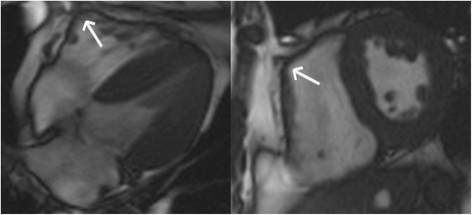
Misdiagnosis of ARVC - Axial and short-axis bright blood images in a control subject. Note the “tethering” of the mid right ventricular free wall to the anterior chest wall (arrows), giving the right ventricle a dyskinetic appearance.
Official websites use .gov
A
.gov website belongs to an official
government organization in the United States.
Secure .gov websites use HTTPS
A lock (
) or https:// means you've safely
connected to the .gov website. Share sensitive
information only on official, secure websites.
Misdiagnosis of ARVC - Axial and short-axis bright blood images in a control subject. Note the “tethering” of the mid right ventricular free wall to the anterior chest wall (arrows), giving the right ventricle a dyskinetic appearance.