
Figure 3. Effect of MC-LR on Leydig cell viability after 48 h incubation.
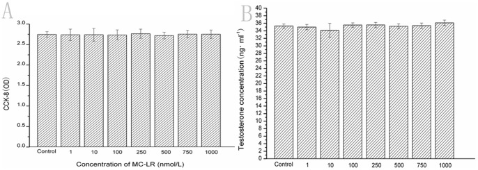
2×10⁵ Leydig cells in 100 µl culture medium were plated in 96-well plates followed 12 hours later by addition of MC-LR at the final concentration of 0, 1.0, 10.0, 100.0, 250.0, 500.0, 750.0 and 1000.0 nmol/L. 48 h later, the effect of MC-LR on cell viability was determined by the CCK-8 assay as described in Materials and Methods. Results represent the means ± SEM of six samples. Effect of MC-LR Testosterone levels produced by Leydig cells in vitro. Leydig cells were cultured with MC-LR at the final concentration of 0, 1.0, 10.0, 100.0, 250.0, 500.0, 750.0 and 1000.0 nmol/L for 48 hours, then the supernatants were collected to analyze the testosterone levels by using an ELISA kit. Results represent the means ± SEM of three samples.