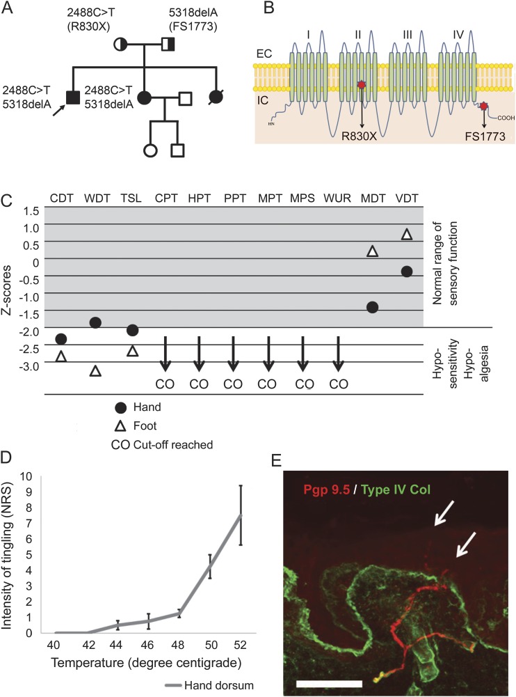
Figure. Detailed sensory phenotyping in a patient with compound heterozygous null mutations in Nav1.7.
(A) The family pedigree shows the proband (arrow) and 2 sisters with congenital inability to experience pain. (B) Cartoon shows the structure of Nav1.7 showing the typical 4-domain structure of voltage-gated sodium channels each with 6 transmembrane segments. The mutations are as follows: a premature stop codon at arginine 830 (within domain 2) and a frameshift at glutamate 1773 within the C-terminal domain. (C) Graphical representation of sensory testing in the proband expressed as Z scores: cold detection threshold (CDT), warm detection threshold (WDT), thermal sensory limen (TSL), cold pain threshold (CPT), heat pain threshold (HPT), pressure pain threshold (PPT), mechanical pain threshold (MPT), mechanical pain sensitivity (MPS), wind-up ratio (WUR), mechanical detection threshold (MDT), and vibration detection threshold (VDT). (D) Graph represents the patient's rating of his tingling sensation on a numerical rating scale (means ± SE) in response to suprathreshold thermal stimuli delivered in a randomized manner. The threshold for this distinct sensation is clearly in the noxious range and the intensity of the sensation encodes the strength of the thermal stimulus. (E) Photomicrograph of skin section immunostained for PGP 9.5 (red) to demonstrate nerve fibers and collagen type IV (green) to show the basement membrane. Fibers are clearly seen in the dermal plexus and some are crossing into the epidermis (arrows); however, the intraepidermal nerve fiber densityis reduced to 3.98 fibers/mm. Scale bar: 50 µm.

