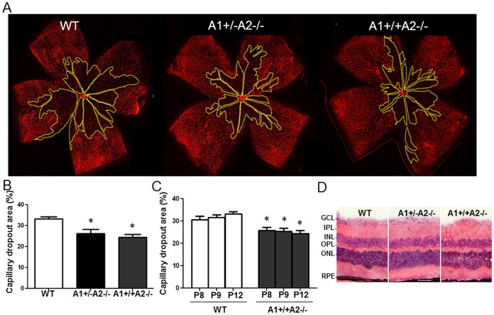
Figure 2. Arginase 2 deletion limits hyperoxia-induced retinal vaso-obliteration.
Wild type (WT), arginase 2-deficient mice (A1+/+A2−/−) and arginase-deficient mice lacking one copy of arginase 1 (A1+/−A2−/−) were placed in 70% oxygen on P7 and prepared for analysis on P8, P9 or P12. Retinal vessels were visualized by lectin labeling (A) and the area of capillary dropout (yellow) was quantified in fluorescence micrographs using ImageJ (B, C). n = 5–9, *P≤0.05 vs WT. Images of hematoxylin and eosin stained cryostat sections from adult mice show comparable retinal morphology in WT, A2−/− and A1+/−A2−/− retinas (D, GCL: ganglion cell layer, IPL: inner plexiform layer, INL: inner nuclear layer, OPL: outer plexiform layer, ONL: outer nuclear layer, RPE: retinal pigment epithelium, scale bar = 50 µm).

