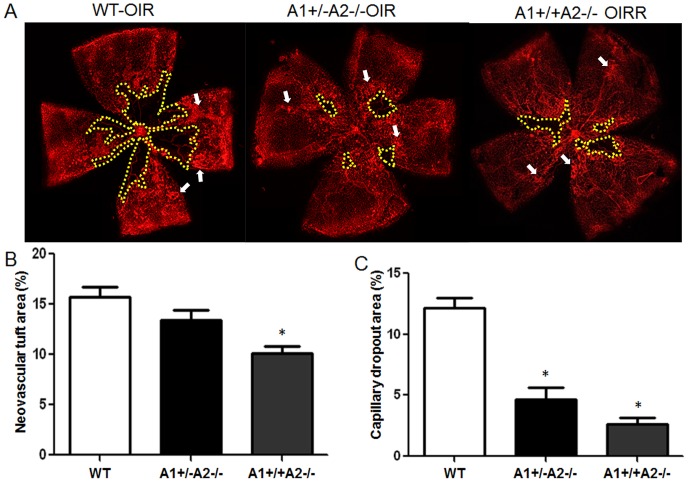
Figure 3. Arginase 2 deletion limits pathological vitreo-retina neovascularization while enhancing intra-retinal neovascularization.
Wild type (WT), arginase 2-deficient mice (A1+/+A2−/−) and arginase 2-deficient mice lacking one copy of arginase 1 (A1+/−A2−/−) were maintained in 70% oxygen from P7 to P12, returned to normoxia for 5 days and prepared for analysis on P17. Retinal vessels were visualized by lectin labeling (A) and areas of vitreoretinal neovascular tufts (arrows) and capillary dropout (yellow) were quantified in fluorescence micrographs using ImageJ (B,C). n = 11–13, *P≤0.05 vs WT and A1+/−A2−/− in B and WT in C.

