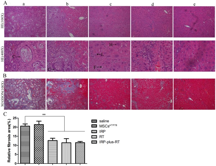
Figure 4. Hematoxylin and eosin and Masson's trichrome staining of liver sections 24 weeks post-transplantation.
A. H&E staining, (a, b): Typical histological finding of cholangiocarcinoma seen in the liver of saline and MSCsATP7B groups. (c–e): Livers of IRP, RT, and IRP-plus-RT rats, respectively. The typical histological finding of cholangiocarcinoma is absent, but the histological finding of chronic hepatitis is present. Enlarged nuclei and fatty changes are visible in some of the hepatocytes; lymphocytes are irregularly distributed among hepatocytes. (C: cholangiocarcinoma; H: regenerated hepatocytes; L: lymphocytes; F: fine fatty droplets); B. MT staining. C. Quantitation of MT staining. The liver fibrosis of IRP, RT, and IRP-plus-RT groups are compared with the saline group, respectively; IRP and RT groups are also compared with IRP-plus-RT group, respectively. ** p<0.01.

