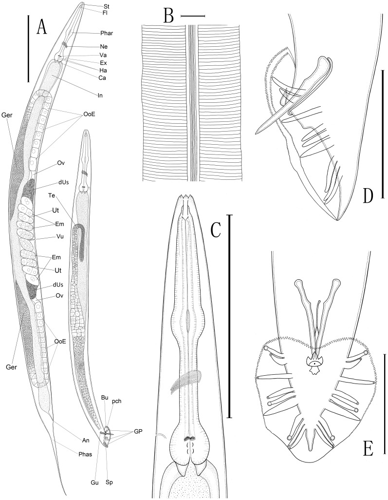
Figure 1. Line drawings of Caenorhabditis sinica sp. n.
A: Overall anatomy of female (left) and male (right)[St: stoma, Fl: flap, Phar: pharynx, Ne: nerve ring, Va: valvular apparatus, Ha: haustrulum, Ex: excretory pore, Ca: cardia (the pharyngo-intestinal valve), In: intestine, Ger: “germigen” containing a well-developed central rachis surrounded by a layer of germ cells, OoE: elongated oocytes, Ov: oviduct, dUs: the distal part of anterior/posterior uterus filled with sperms, Ut: uterus, Em: embryos carried by the uteri, Vu: vulva, An: anus, Phas: phasmid, Te: testis, Bu: bursa, GP: genital papillae, Gu: gubernaculum, Sp: spicule, pch: precloacal hook]; B: Morphology of lateral field (3 ridges flanked by two additional incisures); C: Anterior region of female; D: Lateral view of male caudal region; E: Ventral view of male caudal region with bursa and genital papillae. (Scale bars: A = 200 µm; B = 10 µm; C = 150 µm; D, E = 50 µm).

