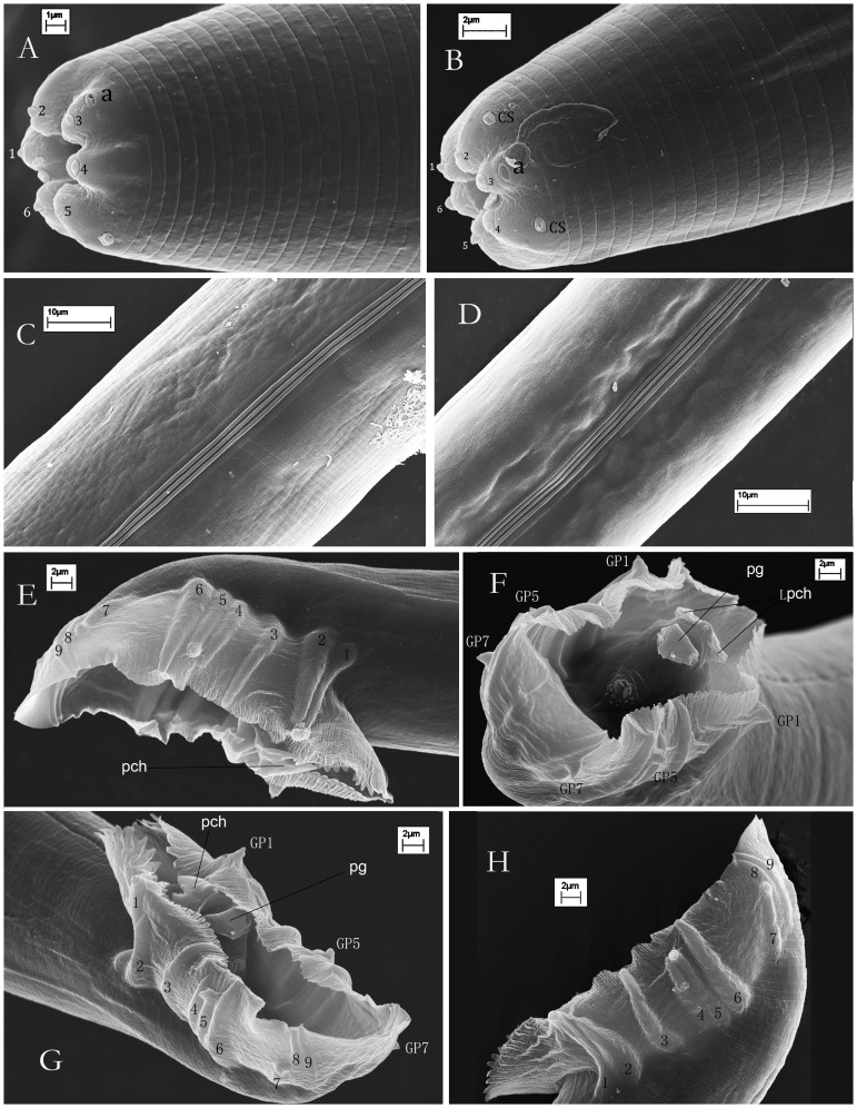
Figure 3. Scanning electron micrographs of Caenorhabditis sinica sp. n.
A, B: Anterior regions of female (A) and male (1–6: six labial sensilla; a: amphid; CS: cephalic sensilla, which are present only in males); C: Lateral field of female; D: Lateral field of male; E, F, G, H: Male caudal regions enveloped by a closed bursa and its nine pairs of genital papillae (pch: precloacal hook; Lpch: the two lateral points of the precloacal hook; pg: posterior part of gubernaculum; GP1, GP5, GP7: showing their dorsal tips being of typical papilliform sensilla).

