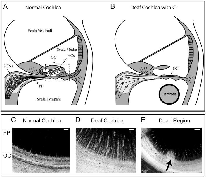
Figure 1.
Upper panel; Schematic diagram of a cross-section through a normal cochlea (A) showing the three fluid filled compartments of the cochlea – the scalae vestibuli, media and tympani. The sensory hair cells (HCs) are located in the organ of Corti (OC) and the cell bodies of the spiral ganglion neurons (SGNs) are located centrally. The SGN peripheral processes (PP) innervate the sensory HCs. In a deafened cochlea (B), cells within the OC are severely damaged, and a cochlear electrode array can be implanted to electrically activate SGNs. However, the PP continue to retract, ultimately leading to death of the SGNs. Lower panel: surface preparations, providing a top-down view of the OC and PP, show the PP stained black and the OC in a normal hearing guinea pig cochlea (C), a cochlea that has been deafened with aminoglycoside drugs (D), and a deafened cochlea that exhibits a localised region (‘dead region’) of greater loss of SGNs and their PP (E – arrow), (scale bar 20 μm C and D, 40 μm E).

