Figure 3.
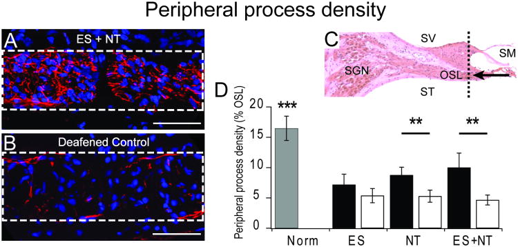
(A and B) example images of cross sections through the osseous spiral lamina (OSL – dotted white box) showing peripheral processes stained with antibodies for neurofilament proteins (red) and cell nuclei stained with DAPI (blue; scale bar 50 μm). Greater process density was observed in the cochleae that received cell-based NT delivery in combination with chronic electrical stimulation from a CI (ES+NT) compared to the deafened control cochlea. (C) Sections were collected in the periphery of the cochlea (dotted black line) and are viewed from the direction of the arrow (SV – scala vestibuli; SM – scala media; ST – scala tympani; OSL – osseous spiral lamina). (D) Means (± standard errors) for peripheral process density within the OSL (dotted box in A and B) of normal hearing cochleae (grey bar), treated cochleae (black bars) and the untreated deaf, contralateral control cochleae (white bars) for each experimental cohort. NT treatment resulted in significantly greater density of peripheral processes compared to the deafened control cochleae. The effect was enhanced with chronic electrical stimulation (NT + ES). There was no effect of electrical stimulation on its own (ES).

