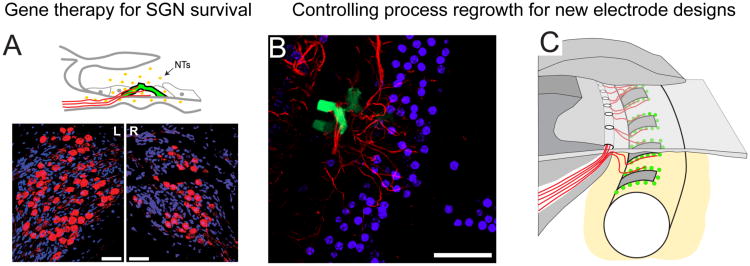
Figure 5.
(A) Schematic diagram illustrating supporting cells (pillar cells) transfected with NT genes (green GFP labelled) in the ototoxically damaged organ of Corti. The transfected cells produce and release NTs (yellow dots) that are accessible to the SGNs and their peripheral processes (red). Lower panels: NT gene therapy resulted in greater SGN survival in the treated (L) cochlea compared to the untreated contralateral cochlea (R), as indicated by a significantly greater density of SGNs in Rosenthal's canal (scale bar 50 μm). (B) A transfected pillar cell (green) in the organ of Corti (top down view). The peripheral processes (red) grew towards cells transfected with NT genes. There was a significantly greater density of processes around NT expressing cells compared to cells expressing the control (GFP only) vector. (C) Schematic illustration depicting the potential of using cell or gene therapy to provide localised sources of NTs in order to promote SGN survival and attract regrowing peripheral processes in a controlled manner. An electrode with this capability might have a greater density of electrode contacts to enable higher resolution and provide greater pitch and temporal information. (Figure 5A adapted from [62] and Figure 5B from [58]).

