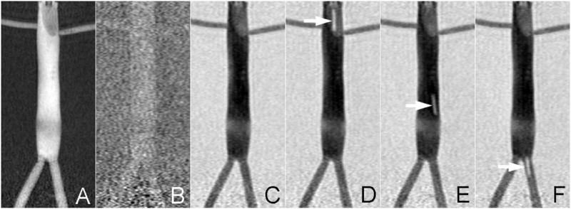
Figure 2.

The roadmapping mode is demonstrated on a vascular phantom that mimics the distal aorta. IA contrast is administered at the start of the run to highlight the vasculature (A). This reference is subtracted from all subsequent scans and thus initially there is minimal contrast (B). The roadmap only appears once the contrast injection is washed out (C) and is durable for the remainder of the run. It is possible to track bright objects against this roadmap as demonstrated by inflating the balloon catheter with 1mM Gd (arrows in D-F).
