
Figure 3.
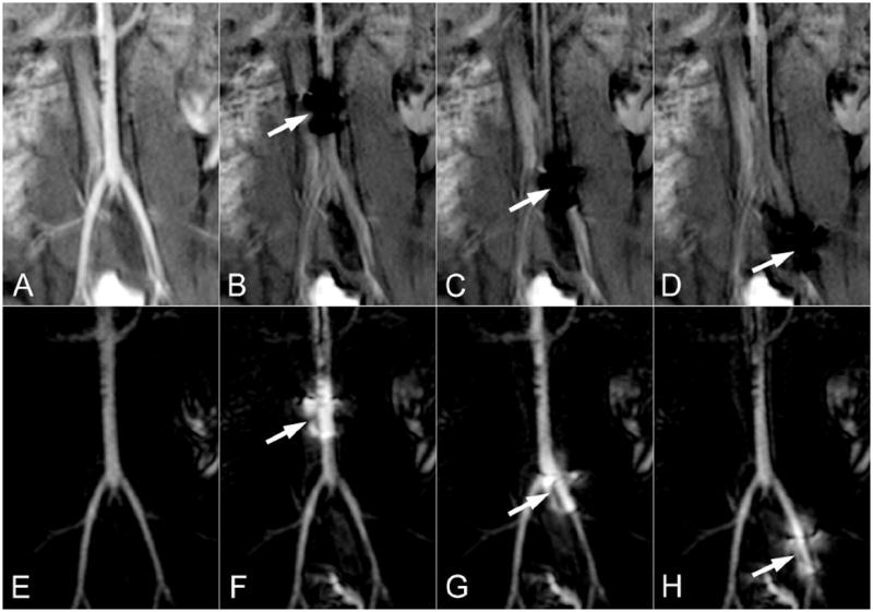
The roadmapping approach is demonstrated in the distal aorta of a swine. The source images are shown on the top row and the roadmapping mode images are on the bottom row. An IA injection of Gd is initially used to highlight arterial anatomy (A) and establish the roadmap (E). The MARC catheter is then activated, producing a substantial signal void on the source images (arrows in B-D) and a signal enhancement pattern on the roadmap images that is superimposed on the arterial anatomy (arrows in F-H). The roadmap images can be used to track the device without losing visibility of the local arterial anatomy.