
Figure 4. Representative microphotographs of urethral cross section at stricture site (stained by Sirius red).
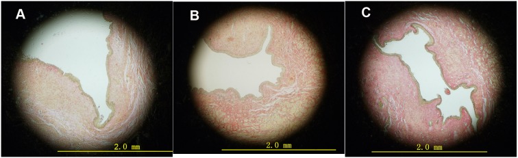
A Urethral cross section of rabbit in DH group. Note a large urethral lumen with light submucosa collagen staining. B Urethral cross section of rabbit in DL group. Note large urethral lumen with light submucosa collagen staining. C Urethral cross section of rabbit in C group. Note almost atresic urethra with deep collagen staining.