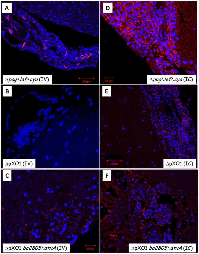
Figure 6. Immuno- histopathology of meninges from rabbits that were infected IV or IC with different mutants.
Immuno-staining of tissues (parallel to figure 5) using anti- killed capsular bacteria anti-serum (Atto 594 – red) and cell nuclei (DAPI – blue). Strain genotype and infection route are as indicated. B. anthracis were observed and demonstrated by immunodetection with all pathogenic strains (A, C-F), but was not present with the non-pathogenic mutant, ΔpOX1 (panel B). Scale bar is 20–50 µm as indicated.

