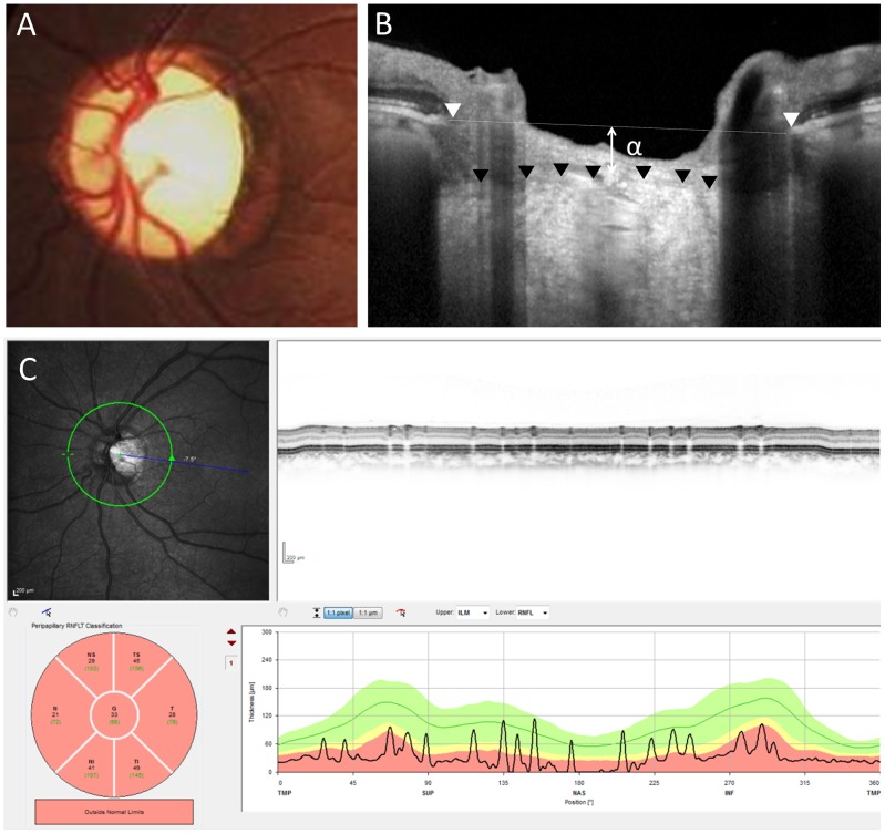
Figure 3. Representative case of eye with compressive optic neuropathy caused by a meningioma.
A fundus photograph (A) shows optic nerve atrophy with pallor and enlarged optic disc cupping. A vertical OCT (B) scan shows a distance of 165 µm between Bruch's membrane opening reference line and the anterior lamina cribrosa (α). A circular OCT (C) scan shows severe thinning of the circumpapillary retinal nerve fiber layer (33 µm).

