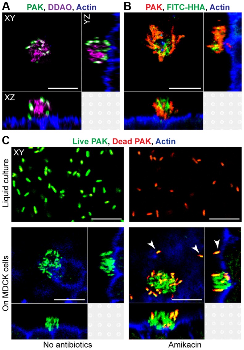
Figure 1. Cell-associated aggregates show key characteristics of biofilms.
(A) MDCK cells were infected with PAK-GFP (green), fixed, and stained for actin (blue) and extracellular DNA with DDAO (purple). (B) MDCK cells were infected with PAK-mCherry (red), fixed, and stained for actin (blue) and with FITC-HHA, which binds to Psl. (C) Bacterial viability after exposure to amikacin was evaluated by staining live bacteria with SYTO 9 (green) and counterstaining dead bacteria with propidium iodide (red). MDCK cells were also stained for actin (blue). Bacteria were incubated in liquid culture (MEM, top row) or with MDCK cells (bottom row). After 60 minutes of infection, bacteria in liquid culture or on MDCK cells were treated with amikacin (400 ug/ml) for 2 hours (top right and bottom right panels). Adherent single bacteria showed propidium iodide staining after amikacin treatment (white arrows). Representative confocal images from three independent experiments are shown. Scale bars, 10 µm.

