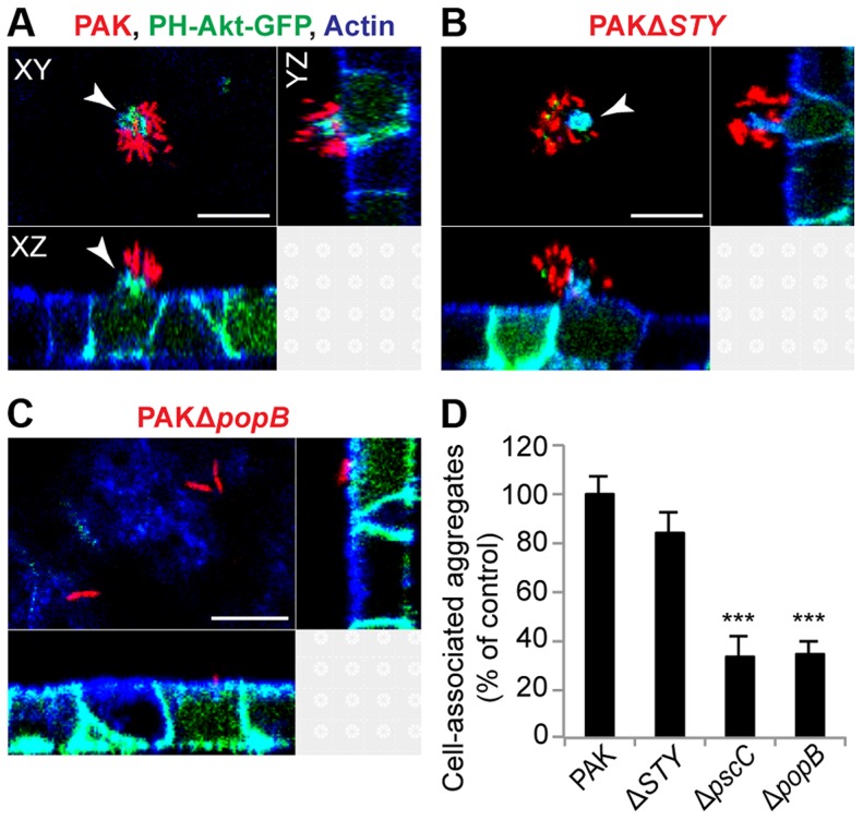
Figure 2. Cell-associated aggregation requires the T3SS translocon but does not require T3SS effectors.
MDCK cells stably transfected with PH-Akt-GFP (green) were infected with mCherry-expressing (A) PAK, (B) PAKΔSTY (lacks the known T3SS effectors), or (C) PAKΔpopB (has the functional needle apparatus but lacks the translocon) for 60 minutes, fixed, and stained for actin (blue). Cell-associated aggregates (red) were visible with PAK (A) and PAKΔSTY (B) and were accompanied by formation of membranous protrusions (white arrows) containing PH-Akt-GFP. PAKΔpopB (C) bound individually or as groups of 2 to 3 bacteria. Representative confocal images from three independent experiments are shown. Scale bars, 10 µm. (D) Cell-associated aggregation by PAK and T3SS mutants was quantified using spinning disk confocal microscopy. Shown is the number of aggregates (≥10 bacteria) normalized to PAK (n≥3 independent experiments). Data are mean ± SEM. ***p<0.001 compared to PAK. Statistics in Supplemental Statistical Analysis (Text S1).

