
Figure 7.
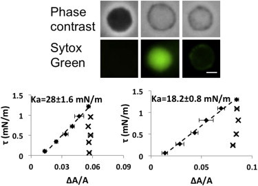
(Top) Paired images of spheroplasts in phase contrast and in the green channel to detect the nucleic acid stain of Sytox Green (at 1 μM): 1st column, the spheroplast with a strong phase contrast showed little penetration by Sytox Green; 2nd column, a spheroplast without interior phase contrast showing very bright Sytox Green in the interior; 3rd column, a spheroplast without interior phase contrast had Sytox Green only around the periphery indicating that the Sytox Green had leaked out. (Bottom) Aspiration experiments on spheroplasts without interior phase contrast (showing two examples). Once the membranes were stretched (diamonds), the membrane area could not recover the low tension value when the tension was reduced (crosses). To see this figure in color, go online.