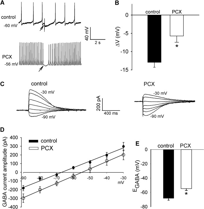
FIGURE 1.
Paclitaxel treatment diminishes GABA-mediated synaptic inhibition and causes a depolarizing shift in EGABA in spinal dorsal horn neurons. A and B, representative perforated recordings and mean data show changes in membrane potentials in response to puff application of 100 μm GABA to lamina II neurons in vehicle-treated control and paclitaxel (PCX)-treated rats (n = 16 and 18 neurons, respectively). Arrows indicate the time when GABA was puffed to the neuron. C and D, original current traces and mean current-voltage plot data show the GABA-elicited currents recorded at different holding potentials in lamina II neurons from vehicle-treated control and paclitaxel-treated rats (n = 18 and 19 neurons, respectively). E, mean data show the difference in the GABA reversal potentials of lamina II neurons recorded in D. *, p < 0.05 compared with the control group. Error bars represent the S.E.

