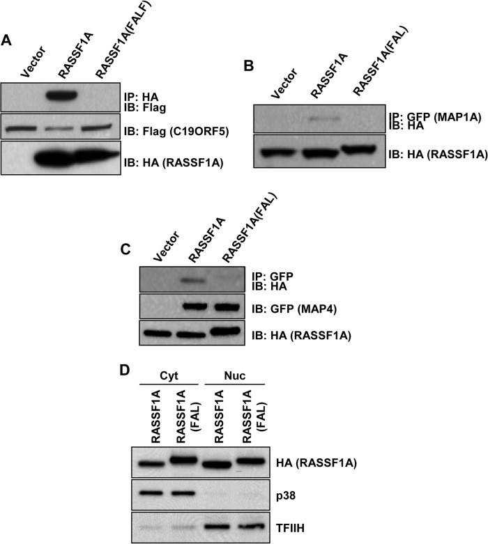
FIGURE 3.
A–C, HEK-293T cells were cotransfected with HA-tagged RASSF1A wild-type or the FAL mutant and FLAG-tagged C19ORF5 (A), GFP-tagged MAP1A (B), or GFP-tagged MAP4 (C), and equal amounts of protein were immunoprecipitated (IP) with anti-HA (A) or anti-GFP (B and C) antibodies. The immunoprecipitates were fractionated on SDS gels and Western blotted with anti-FLAG, anti-HA, and anti-GFP antibodies. Levels of MAP1A were not measured by Western blot analysis because the size of MAP1A precluded it from entering the gel. However, the intensity of GFP was the same for each transfection, as determined by fluorescence microscopy. IB, immunoblot. D, HEK-293T cells were transfected with HA-tagged wild-type RASSF1A or RASSF1A(FAL). Cells were lysed, and cytoplasmic (Cyt) and nuclear (Nuc) fractions were prepared and analyzed by Western blotting using an anti-HA antibody. p38 and TFIIH were used as markers for the cytoplasmic and nuclear fractions, respectively.

