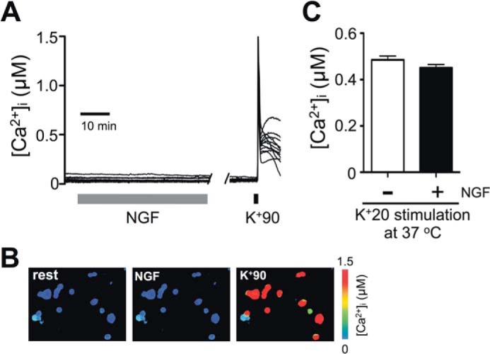
FIGURE 2.

NGF does not affect [Ca2+]i in DRG neurons. Cultured DRG neurons were prepared using the protocol described in Fig. 1A. A, representative traces of [Ca2+]i from Fura-2/AM-loaded DRG neurons during treatment with 25 ng/ml NGF (gray bar). Cell viability and responsiveness to depolarization were confirmed at the end of the experiment by applying 90 mm KCl (30 s) to the cells. Ca2+ imaging was performed at room temperature (n = 64 cells, 4 independent experiments). Each trace represents an individual cell. B, [Ca2+]i in cells is indicated by pseudocolor images taken at rest (left), during the NGF treatment (middle) and at the peak of K+90-induced depolarization (right) for the same experiment as described in A. C, comparison of the effects of prolonged (6-h) K+20 depolarization on [Ca2+]i in the absence (−NGF; white) or presence (+NGF; black) of 25 ng/ml NGF. The K+20 solutions were supplemented with 1 μm BayK8644. [Ca2+]i was measured at the end of the 6-h treatment, in cells loaded with Fura-2/AM. All procedures were conducted at 37 °C. p > 0.05, Student's t test; n = 99 cells (−NGF) and 121 cells (+NGF); 3 independent experiments/condition. All data are presented as mean ± S.E. (error bars).
