
FIGURE 9.
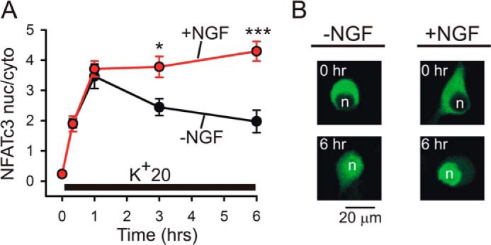
NGF increases depolarization-induced nuclear translocation of NFATc3 in DRG neurons. A, DRG neurons transfected with EGFP-NFATc3 were deprived of NGF for 24 h prior to stimulation. K+20 (supplemented with 1 μm BayK8644) was applied in the absence (black) or presence (red) of 25 ng/ml NGF under conditions similar to those described for the NFAT-luciferase experiments (Figs. 1 and 4–6). The translocation of EGFP-NFATc3 was imaged and quantified as described previously (33). In the absence of NGF, EGFP-NFATc3 was gradually exported from the nucleus despite continuous depolarization, whereas in NGF-treated DRG neurons, the levels of EGFP-NFATc3 in the nucleus continued to increase slowly throughout the period of depolarization. *, p < 0.05; ***, p < 0.001, one-way ANOVA with Bonferroni's post hoc test (12–47 cells). Data are presented as mean ± S.E. (error bars). B, representative images showing distribution of EGFP-NFATc3 in DRG neurons at rest and after 6 h of K+20 stimulation in the absence (left) or presence (right) of 25 ng/ml NGF. n, cellular nuclei.