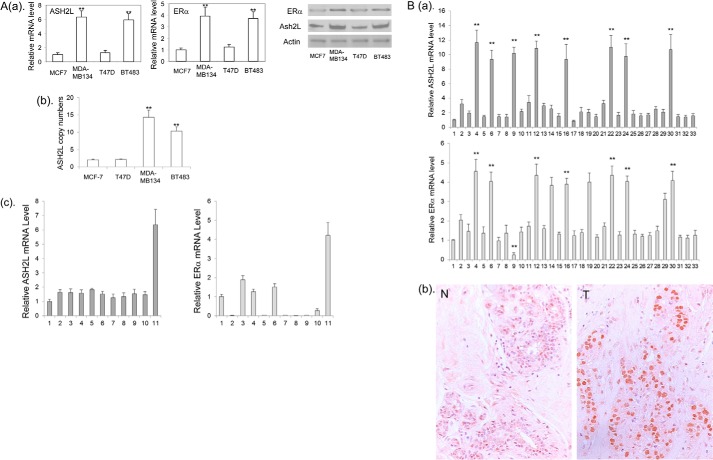
FIGURE 1.
A: a, the levels of ERα expression in breast cancer cell lines are correlated with the levels of ASH2L expression. The ERα and ASH2L mRNA (left panel) and protein (right panel) in MCF-7, T47D, BT483, and MDA-MB-134 cells were determined by real-time RT-PCR and Western blot, respectively. **, p < 0.01 (versus MCF-7 and T47D). b, copy number of the ASHL gene was determined in MCF-7, T47D, BT483, and MDA-MB-134 cells by real-time PCR. **, p < 0.01 (versus MCF-7 and T47D). c, ASH2L and ERα expression in additional breast cancer cell lines. Lanes 1, primary breast epithelial cells; 2, MCF-10A; 3, MDA-MB-361; 4, ZNF-75–1; 5, BT-20; 6, BT474; 7, SKBR3; 8, MDA-MB-231; 9, MDA-MB-157; 10, MDA-MB-175; 11, MDA-MB-134. B: a, real-time PCR analyses of ASH2L and ERα mRNA in human primary ER positive breast cancers. Lanes 1, human normal mammary epithelial cells served as a normal control; 2 to 33, primary breast cancers. **, p < 0.01 (versus other tumors). b, immunostaining of ASH2L protein. N, normal breast; T, breast cancer with increased expression of ASH2L protein (tumor number 4 from a is shown as a representative).

