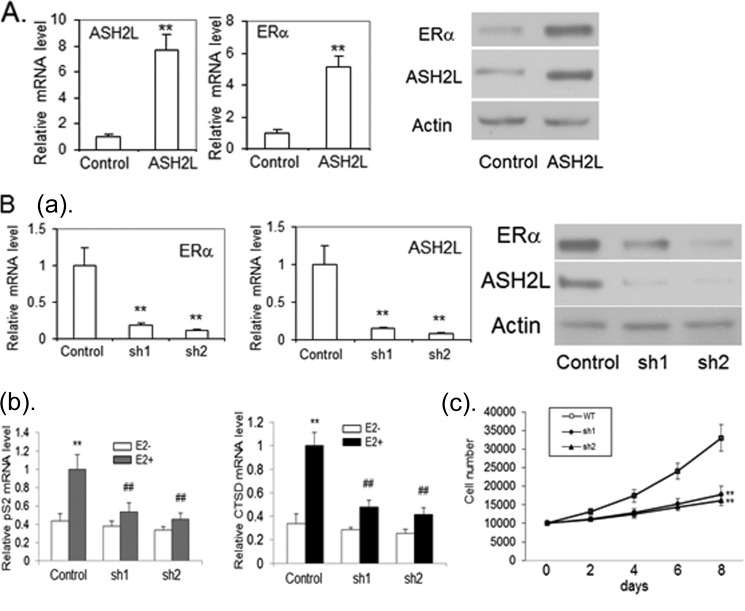
FIGURE 2.
A, ASH2L induces ERα expression in mammary epithelial cells. Immortalized mammary epithelial cells were infected with lentivirus expressing ASH2L or LacZ as a control. The ASH2L and ERα mRNA were examined by real-time RT-PCR, which were normalized to control β-actin mRNA levels (left panel). **, p < 0.01 (versus control). The ASH2L and ERα protein were examined by Western blot (right panel). B: a, depletion of ASH2L leads to deceased expression of ERα in human breast cancer cells. BT483 cells were infected with viruses expressing two different shRNAs targeting ASH2L (sh1, sh2) or control shRNA. Real-time RT-PCR was performed to determine ERα and ASH2L mRNA levels (left panel). **, p < 0.01 (versus control). The level of ERα and ASH2L protein were evaluated by Western blot (right panel). b, reduced expression of ERα target genes with the depletion of ASH2L expression. The cells were treated with 100 nm E2 or control vehicle for 24 h and collected for the preparation of total RNA. Real time RT-PCR was performed to determine cathepsin D (CTSD) and pS2 mRNA levels, which were normalized to internal control β-actin mRNA levels. **, p < 0.01 (versus control vehicle). ##, p < 0.01(versus control shRNA). c, depletion of ASH2L results in cell growth inhibition of BT483. ASH2L-depleted cells (sh1 and sh2) and cells expressing control shRNA (WT) were counted by trypan blue staining at different times after initial seeding of 2 × 10⁴ cells with the treatment of 100 nm E2. The experiment was done independently three times, and the results were averaged with error bars representing S.D. **, p < 0.01 (versus control shRNA).

