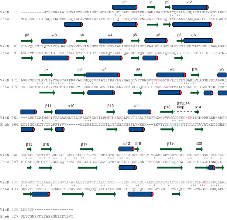
FIGURE 3.
Amino acid sequence comparison of VinN with PheA. Specificity-conferring code residues are shown in red. The residues involved in AMP binding in PheA are shown in blue. The secondary structural elements of VinNN and PheA are indicated by bars above or below the sequence. The VinN β13β14 loop containing Lys330 and Arg331 is indicated by a broken line above the sequence. The C-terminal domain sequence of VinN is shown in gray. The conserved positions are marked with an asterisk.

