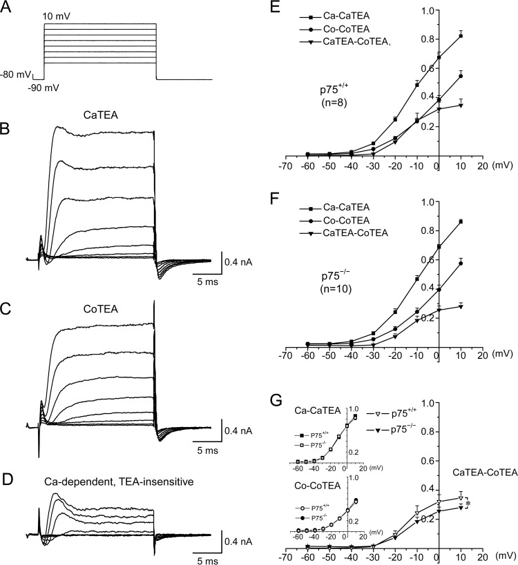
FIGURE 2.
p75 facilitates Ca2+-dependent, TEA-insensitive K+ current in Purkinje cells. A, voltage steps used for whole-cell voltage clamp recording of acutely dissociated Purkinje cells. B and C, representative traces from a wild type neuron bathed in the CaTEA solution containing 2 mm Ca2+, 300 nm TTX, and 1 mm TEA (B) or bathed in the CoTEA solution containing 2 mm Co2+, 300 nm TTX, and 1 mm TEA (C). D, subtracted traces of B and C, representing the activity of Ca2+-dependent but TEA-insensitive K+ channels. The inward currents observed at the start of the recording represent calcium channel activities. E and F, average current-voltage (I-V) curves under the conditions depicted in B–D for p75+/+ (E) and p75−/− (F) Purkinje neurons. G, comparison of I-V curves between Purkinje cells from p75+/+ and p75−/− mice revealed that Ca2+-dependent and TEA-insensitive K+ currents were reduced by 23% in p75−/− Purkinje cells as compared with the wild type. Note that in E–G, current amplitudes were normalized to the maximal value obtained in the absence of TEA but in the presence of 2 mm Ca2+; thus, the y axis in E–G indicates normalized currents. Significant differences (analysis of variance; p < 0.05) are found for the Ca2+-dependent TEA-insensitive K+ currents (CaTEA-CoTEA) between −30 and 10 mV. Error bars, S.E.

