
FIGURE 2.
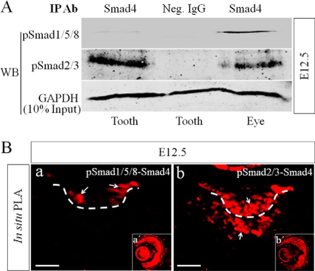
Lack of pSmad1/5/8-Smad4 complex but abundant pSmad2/3-Smad4 complexes are present in early dental mesenchyme. A, Western blot assay shows barely detectable pSmad1/5/8-Smad4 complexes but abundant pSmad2/3-Smad4 complexes in the precipitated immunocomplexes from E12.5 tooth germs. E12.5 eye samples were included as positive controls. IP Ab, immunoprecipitation antibody; Neg. IgG, negative control IgG; WB, Western blot. B, in situ PLA shows the presence of a small amount of pSmad1/5/8-Smad4 complexes in the dental epithelium (panel a) and abundant pSmad2/3-Smad4 (panel b) complexes in E12.5 tooth germ. Eyes were used as positive controls (panels a′ and b′). White arrows point to PLA signals (red fluorescence). Bar = 50 μm.